Everything you need to know about fibroids — what they are, why they grow, the different types, how they're found, and what modern treatment looks like. Explained simply, without the medical jargon.
Uterine fibroids (also called leiomyomas or myomas) are noncancerous (benign) growths that develop in or around the muscular wall of the uterus. They are made of smooth muscle cells and fibrous connective tissue.
Despite their name, fibroids are not made of fibrous tissue — the name is somewhat misleading. What matters most is that they are almost always benign (non-cancerous). Less than 1 in 1,000 fibroid cases is cancerous. This means you don't need to fear a cancer diagnosis — but you do need to treat the symptoms they cause.
Fibroids can range in size from as small as a pea to as large as a watermelon. A single woman may have just one fibroid or many. They may stay the same size for years or grow rapidly. Their behavior is largely driven by the hormone estrogen.
The exact cause of fibroids is not fully understood, but researchers have identified several key factors that contribute to their development and growth.
Estrogen and progesterone — the hormones that regulate your menstrual cycle — appear to promote fibroid growth. Fibroids typically shrink after menopause when these hormones decline. This is why hormonal birth control can sometimes temporarily reduce symptoms.
Fibroids tend to run in families. If your mother, sister, or maternal grandmother had fibroids, your risk is significantly higher. Certain genetic mutations are linked to fibroid development, though the specific genes involved are still being studied.
Black women are 2–3 times more likely to develop fibroids than white women, tend to develop them at a younger age, and often experience more severe symptoms. Hispanic women also have higher rates than white women. Researchers believe both genetic and environmental factors contribute to these disparities.
Fibroids are most common during the reproductive years (ages 30–50), when estrogen levels are highest. They are rare before puberty and typically shrink after menopause. However, some women in their 20s do develop symptomatic fibroids, particularly Black women.
A diet high in red meat and low in fruits and vegetables may increase fibroid risk. Obesity (excess body fat produces extra estrogen), alcohol consumption, and vitamin D deficiency have also been linked to higher fibroid rates. Some studies suggest green vegetables and citrus fruits may be protective.
Women who have never given birth (nulliparous women) have a higher risk of developing fibroids. Pregnancy temporarily reduces estrogen exposure and may offer some protection. However, pregnancy itself can sometimes accelerate fibroid growth due to hormonal changes.
Not all fibroids are the same. Their location within or around the uterus plays a major role in what symptoms you experience and how severe they are.
These fibroids grow inside the uterine cavity, just beneath the inner lining (endometrium). Even small submucosal fibroids can cause severe bleeding and fertility problems because they disrupt the uterine lining.
Common symptoms: Very heavy bleeding, prolonged periods, large blood clots, anemia, difficulty conceiving, miscarriage.
These are found within the muscular wall of the uterus (myometrium) — the most common type. As they grow, they can expand and distort both the uterine cavity and its outer surface.
Common symptoms: Heavy bleeding, pelvic pain and pressure, enlarged uterus, back pain, urinary or bowel symptoms as they grow.
These grow on the outer surface of the uterus, projecting outward into the pelvic cavity. They tend to cause more pressure symptoms than bleeding because they don't directly affect the uterine lining.
Common symptoms: Pelvic pressure, back pain, frequent urination, bloating, pain during sex, leg pain.
These fibroids are attached to the uterus by a narrow stalk (like a mushroom). They can be pedunculated intracavitary (inside) or pedunculated subserosal (outside). If the stalk twists (torsion), it can cause sudden, severe pain.
Common symptoms: Vary by location — pressure, pain, or bleeding. Sudden severe pain if torsion occurs.
Many women don't know they have fibroids until a routine pelvic exam or ultrasound reveals them. Here are the most common ways fibroids are detected and evaluated.
During a routine gynecological exam, your doctor may feel an enlarged or irregularly shaped uterus, suggesting the presence of fibroids. This is often the first clue that fibroids exist.
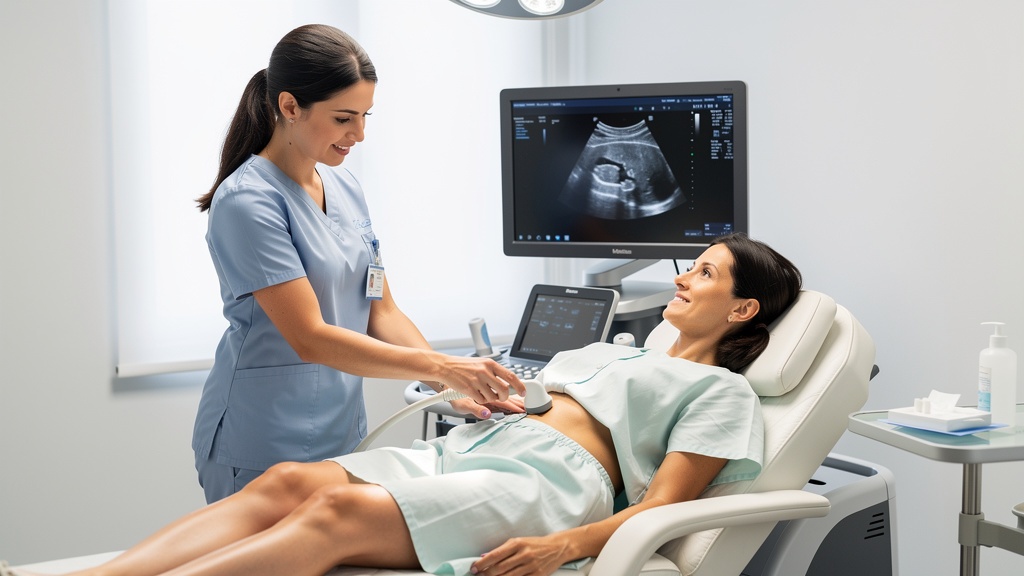
The most common first-line imaging tool. A transabdominal or transvaginal ultrasound uses sound waves to create images of your uterus and can confirm the presence, size, number, and location of fibroids.
The gold standard for fibroid evaluation. An MRI provides detailed images of each fibroid's exact size, location, and blood supply. It is essential for planning UFE and ensures the most accurate, effective treatment possible.
Saline is gently introduced into the uterus during an ultrasound to get a clearer view of the uterine cavity. Particularly useful for detecting submucosal fibroids that may cause heavy bleeding.
A thin, lighted camera is inserted through the cervix to view the inside of the uterus. This can diagnose submucosal fibroids and rule out other conditions like polyps or endometrial cancer.
A complete blood count (CBC) can check for anemia caused by heavy fibroid bleeding. Other blood tests may check kidney function, thyroid levels, or rule out bleeding disorders that could explain heavy periods.
Knowledge is power — but knowledge without action is just worry. If you recognize fibroid symptoms in your life, here's what to do next:
Heavy bleeding, pelvic pain, and constant fatigue are not things you have to simply endure. These are medical conditions with effective treatments.
In just 5 minutes, our short questionnaire will help you understand whether UFE is likely a good fit for your situation.
Our UFE specialist will review your symptoms, imaging results, and health history to give you a clear, personalized recommendation — with no obligation.
If another doctor has recommended hysterectomy or myomectomy, a second opinion from an interventional radiologist who specializes in UFE is always worthwhile. You may have more options than you were told.